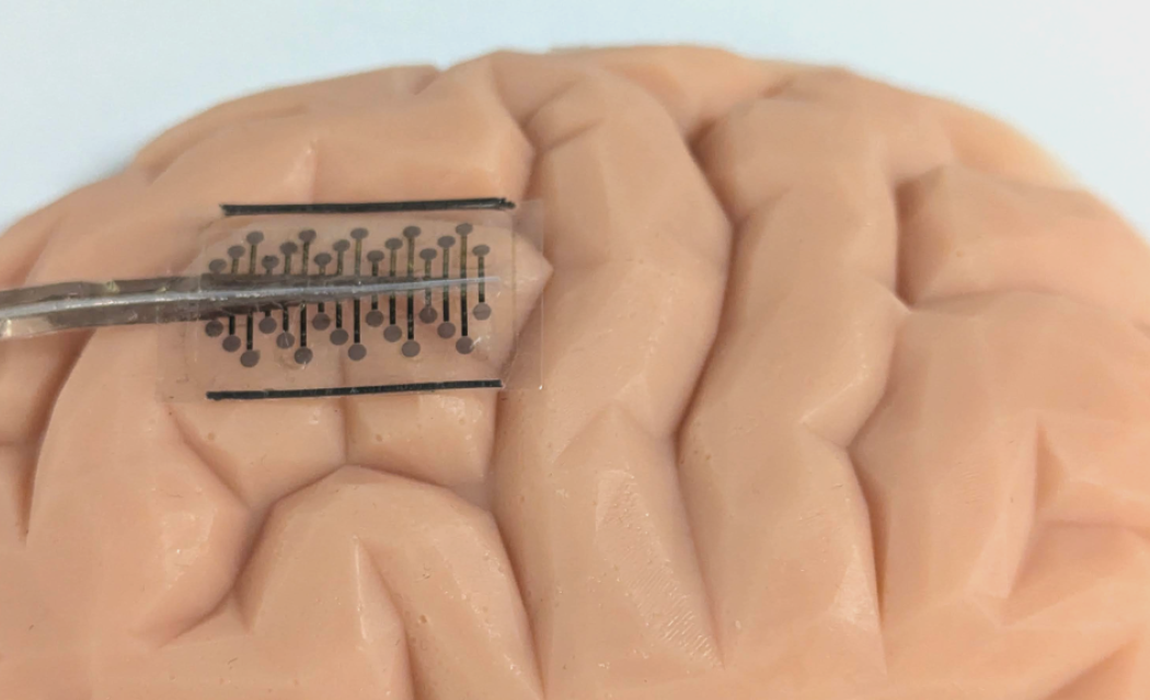
A research team led by the University of Oxford and the University of Cambridge have created new ‘origami-inspired’ brain electrodes that can fold up to a fraction of their full size, potentially significantly reducing the amount of surgery needed to treat conditions such as epilepsy, or to install brain-computer interfaces.
Measuring brain electrical activity is essential to diagnose and treat conditions such as epilepsy. However, this often requires surgeons to cut out a large window in the skull (a craniotomy) to place electrodes directly onto the brain surface. This procedure typically entails a prolonged recovery period and poses severe infection risks. The new study, published in Nature Communications, demonstrated that using a folding design for brain electrodes could reduce the incision area needed by about five times, without affecting functionality.
Senior author Associate Professor Christopher Proctor (Department of Engineering Science, University of Oxford) said: “This study presents a new approach to directly interfacing with large areas of the brain through a key-hole like surgery. The potential significance of this work is two-fold. First, there is the promise of a less invasive diagnostic tool for epilepsy patients. Second, we envision the minimally invasive nature will enable new applications in brain machine interfaces.”
When fully expanded, the device resembles a flat, rectangular silicone wafer with 32 embedded electrodes, attached to a cable. The 70 microns thick wafer is folded up, enabling it to fit through a slit just 6 mm across. Once in position on the brain surface, a pressurised fluid-filled chamber in the wafer inflates and unfolds the device to cover an area five times larger, up to 600 mm2.
According to the team, the device could potentially start to be used to treat human patients within a few years. Around 50 million people worldwide have epilepsy, which carries a risk of premature death up to three times higher than for the general population (WHO).
Lead-author Dr Lawrence Coles (Department of Engineering, University of Cambridge) said: “We are now working with clinical partners to refine the design with the aim of starting trials in human patients within two years. Besides epilepsy, this approach could be used to diagnose and treat other conditions that result in brain seizures, such as certain brain tumours.”
According to the researchers, the fold-up design could also reduce the amount of surgery needed to install brain-computer interfaces, which could benefit people with disabilities as well as optimise human-computer interactions.
“Brain-computer interfaces are an incredibly fast-moving and promising area of development” Dr Coles added. “A particularly exciting area is direct speech decoding, where electrodes measure signals directly off the brain surface itself to translate what the person is trying to say.”
Ley Sander, Medical Director at the Epilepsy Society and Professor of Neurology at UCL (who was not involved with the study) said: “This is an exciting new approach to brain surgery, but it is very much in its infancy. Anything that reduces the invasive nature of brain surgery and the risk of infection has to be welcomed, particularly if it promises a shorter recovery time. Brain surgery is only possible for people for whom we are able to pinpoint the area of the brain where their seizures occur, but in these cases, it offers real hope of seizure freedom.”
For more information, please visit the University of Oxford website or Nature Communications who published the study ‘Origami-inspired soft fluidic actuation for minimally invasive large-area electrocorticography’.
Image provided by University of Oxford
